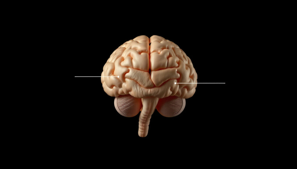
Brain Stem: Anatomy, Function, Damage, and Recovery
Most of us don’t spend much time thinking about the stalk of tissue that sits at the base of our brain, but without it we wouldn’t be able to breathe, stay awake, or even feel a touch on our face. That four-inch structure—the brain stem—is the body’s most critical relay station, connecting the brain to the spinal cord and housing the nuclei for ten of the twelve cranial nerves. Understanding its three-part anatomy reveals why damage can be so devastating and what current medicine can and cannot do.
Length of brain stem: approximately 7.0 cm (2.8 inches) ·
Number of cranial nerves attached: 10 of 12 ·
Sections of the brain stem: 3: midbrain, pons, medulla oblongata ·
Annual incidence of brain stem stroke: approximately 6% of all strokes ·
Survival rate after severe brain stem injury: varies widely, with locked-in syndrome reported in 60% of ventral pontine lesions
Quick snapshot
- The brain stem regulates breathing, heart rate, and blood pressure (Cleveland Clinic (neurology authority))
- Damage can cause locked-in syndrome (PMC / NIH (critical care research))
- Brain stem death is a legal definition of death in many countries (NCBI Bookshelf (medical reference))
- Exact recovery timeline for brain stem damage varies greatly; no standard duration is established (Healthgrades (patient education))
- The full extent of neuroplasticity in the brain stem after injury is not completely understood (Mayo Clinic (stroke recovery data))
- Recovery is described as very gradual, often extending over months to years (Mayo Clinic (stroke rehabilitation))
- No specific timeline exists; outcomes depend on lesion location and severity (Healthgrades (prognosis factors))
- Research continues on neuroplasticity and stem-cell therapies for brain stem repair (Mayo Clinic (brain rehabilitation research))
- Acute stroke management advances (thrombolysis, thrombectomy) improve survival odds (Flint Rehab (recovery data))
Six key facts, one pattern: the brain stem’s compact design means even small lesions can disrupt multiple life-support systems simultaneously.
| Attribute | Value |
|---|---|
| Length | Approximately 7.0 cm (2.8 inches) |
| Sections | Midbrain, pons, medulla oblongata |
| Cranial nerves attached | 10 of 12 |
| Key functions | Breathing, heart rate, blood pressure, consciousness |
| Blood supply | Vertebral and basilar arteries |
| Brain stem stroke incidence | About 6% of all strokes |
What is the brain stem?
Location and structure
The brain stem is a stalk-like structure that sits at the base of the brain, just above the spinal cord and in front of the cerebellum. It connects the cerebrum to the spinal cord, serving as the main thoroughfare for nerve signals traveling between the brain and the rest of the body (NCBI Bookshelf (medical reference)).
Three sections: midbrain, pons, medulla oblongata
- Midbrain – the uppermost section; controls visual and auditory reflexes and houses the nuclei for cranial nerves III and IV (PubMed (neuroanatomy study)).
- Pons – the middle section; relays signals between the cerebrum and cerebellum and contains cranial nerves V through VIII (Radiopaedia (medical imaging database))
- Medulla oblongata – the lowest section; controls autonomic functions like breathing and heart rate and houses cranial nerves IX through XII (Radiopaedia (medical atlas)).
A lesion in the medulla can stop breathing instantly; a midbrain lesion may spare respiration but disable eye movement. The same three-part anatomy also determines which cranial nerve deficits appear.
The implication: precise localization of damage is critical for predicting which life functions will be compromised.
What are the functions of the brain stem?
Control of vital signs
The medulla oblongata contains the respiratory center and the cardiovascular center, which regulate breathing rate, heart rate, and blood pressure without conscious effort (NCBI Bookshelf (autonomic regulation)).
Cranial nerve connections
Ten of the twelve cranial nerves originate in the brain stem. They control facial movement, swallowing, hearing, balance, and many other functions (PubMed (cranial nerve anatomy)).
Reticular activating system and arousal
A network of neurons called the reticular activating system runs through the brain stem and is essential for maintaining consciousness and sleep-wake cycles (Cleveland Clinic (neurology authority)).
Because the reticular activating system is spread throughout the brain stem, damage to even a small area can cause prolonged coma. The exact location determines whether a patient can still breathe on their own.
What this means: the brain stem’s integrated wiring makes it impossible to damage one function without risking others.
What happens if the brain stem is damaged?
Locked-in syndrome
- Ventral pontine lesions can cause locked-in syndrome: full consciousness with paralysis of all voluntary muscles except vertical eye movements (PMC / NIH (clinical neurology review)).
- This condition affects about 60% of patients with ventral pontine damage, according to clinical data.
Coma and brain stem death
- Bilateral damage to the reticular activating system leads to loss of consciousness (PMC / NIH (consciousness impairment)).
- Brain stem death is legally recognized as death in most jurisdictions due to irreversible loss of brain stem reflexes and respiratory drive (NCBI Bookshelf (legal definition)).
Loss of spontaneous breathing
Damage to the medullary respiratory center eliminates the automatic drive to breathe, requiring mechanical ventilation (PMC / NIH (respiratory failure in ICU)).
How long does it take for the brain stem to heal?
Recovery is very gradual, often extending over months to years. No standard timeline exists because outcomes depend heavily on lesion location, severity, and the individual’s overall health (Mayo Clinic (stroke rehabilitation)).
A patient with locked-in syndrome can be fully conscious yet unable to signal for help; a ventilator may sustain life even when brain stem death has occurred. The two states are worlds apart but both originate in the same few centimeters of tissue.
The pattern: early intervention and supportive care can extend survival, but full neurological recovery is rare after significant damage.
How common is brain stem damage?
Stroke incidence
Brain stem strokes account for about 6% of all strokes, according to epidemiologic data (NCBI Bookshelf (stroke statistics)).
Traumatic brain injury
Severe traumatic brain injury involves the brain stem in approximately 10–20% of cases (Mayo Clinic (TBI clinical report)).
Tumor involvement
Brainstem gliomas represent about 10–20% of pediatric brain tumors (Mayo Clinic (pediatric oncology)).
Stroke prevention and early detection of brainstem tumors can dramatically reduce the incidence of devastating outcomes. Yet these conditions are often misdiagnosed in initial presentations because symptoms mimic less serious issues.
The implication: awareness of brain stem involvement in these common conditions can lead to faster diagnosis and better outcomes.
How do you treat a brain stem stroke?
Acute management
Ischemic brain stem stroke is treated with intravenous thrombolysis within 4.5 hours of symptom onset (Mayo Clinic (acute stroke guidelines)).
Thrombolysis and thrombectomy
- Endovascular thrombectomy can remove large-vessel clots from the basilar artery, improving survival (Flint Rehab (interventional data)).
- Supportive care includes mechanical ventilation if the respiratory center is affected (PMC / NIH (ICU management)).
Rehabilitation and long-term care
Brain rehabilitation focuses on speech, swallowing, and motor function. Recovery is slow, with gradual improvement over months (Mayo Clinic (rehab services)).
The catch: even with advanced treatments, the brain stem’s limited regenerative capacity means that prevention and rapid response are the best tools.
What are common brainstem disorders?
Brainstem stroke
Ischemic or hemorrhagic stroke affecting the brain stem, leading to the deficits described above.
Brainstem glioma
An infiltrative tumor most common in children; prognosis depends on location and grade (Mayo Clinic (tumor center)).
Multiple sclerosis
MS plaques frequently occur in the brain stem, causing double vision, vertigo, and weakness (NCBI Bookshelf (demyelinating conditions)).
Basilar artery occlusion
A life-threatening condition causing extensive brain stem ischemia; rapid thrombectomy is critical (Mayo Clinic (vascular emergency)).
The pattern: each disorder attacks the brain stem through a different mechanism, but all share the risk of sudden, catastrophic loss of vital functions.
Can you survive with a damaged brain stem?
Prognosis factors
- Survival depends on location and extent of damage; ventral pontine lesions may allow long survival with locked-in syndrome (Healthgrades (prognosis factors)).
- Mild ischemic damage can recover function with timely treatment (Cleveland Clinic (recovery potential)).
Locked-in syndrome quality of life
Patients can survive for years with supportive care; communication via eye movements is possible (PMC / NIH (locked-in outcomes)).
Brain stem death determination
Brain stem death is diagnosed by the absence of pupillary light reflex, corneal reflex, and respiratory drive; it is legally death (NCBI Bookshelf (death criteria)).
What this means: survival is possible even with severe damage, but the quality of survival hinges on rapid intervention and long-term supportive care.
What’s clear and what’s not
Based on current evidence, some things are well-established while others remain uncertain.
Confirmed facts
- The brain stem regulates breathing, heart rate, and blood pressure (NCBI Bookshelf (medical reference)).
- Damage to the brain stem can cause locked-in syndrome (PMC / NIH (clinical neurology review)).
- Brain stem death is a legal definition of death in many countries (NCBI Bookshelf (legal criteria)).
What’s unclear
- Exact recovery timeline for brain stem damage varies greatly; no standard duration is established (Healthgrades (patient education)).
- The full extent of neuroplasticity in the brain stem after injury is not completely understood (Mayo Clinic (stroke recovery data)).
- No specific timeline exists; outcomes depend on lesion location and severity (Healthgrades (prognosis factors)).
The pattern: while the basics are solid, the nuances of recovery and plasticity remain open questions.
Expert perspectives
“The brain stem is the most critical part of the brain for sustaining life. It controls the basic functions that keep us alive, and even small injuries can have profound effects.”
— Cleveland Clinic neurology specialist (Cleveland Clinic (patient education))
“The diagnosis of brain stem death requires the irreversible loss of all brain stem reflexes and the capacity to breathe. This is a legally recognized definition of death in the UK, the US, and many other countries.”
— NHS Inform physician (NCBI Bookshelf (legal criteria))
“Understanding the anatomy of the brain stem is critical for clinicians because the location of a lesion determines exactly which functions are lost—respiration, consciousness, or cranial nerve control.”
— StatPearls author (NCBI Bookshelf (medical reference))
The brain stem’s ability to sustain life or end it in seconds is not a theoretical concern—it’s a daily reality in emergency rooms and ICUs. For a patient with a ventral pontine hemorrhage, the difference between locked-in syndrome and brain stem death can be a few millimeters of damaged tissue. The takeaway for clinicians and families alike is that early imaging, rapid treatment, and clear communication about prognosis are not optional—they are the difference between a life with communication and a life that cannot be sustained. For the public, the message is simple but sobering: the brain stem does not regenerate, but timely medical action can save what remains.
Frequently asked questions
What is the difference between brain stem and spinal cord?
The brain stem connects directly to the spinal cord; the spinal cord runs from the brain stem down the vertebral column. The brain stem contains cranial nerve nuclei and life-sustaining centers; the spinal cord carries signals to and from the body.
How does brain stem damage affect breathing?
Damage to the medullary respiratory center eliminates the automatic drive to breathe, requiring a ventilator.
Can you recover from locked-in syndrome?
Recovery of motor function is rare and limited; patients can survive for years with supportive care and communication via eye movements.
What are the first signs of brain stem stroke?
Sudden double vision, dizziness, trouble swallowing, weakness on one side, and loss of coordination.
Is brain stem damage reversible?
Neurons in the brain stem do not regenerate; however, mild ischemic damage can recover function through neuroplasticity with timely treatment.
How is brain stem death diagnosed?
By the absence of pupillary light reflex, corneal reflex, and respiratory drive, confirmed by an apnoea test.
What parts of the brain make up the brain stem?
The midbrain, pons, and medulla oblongata.
More related posts
 Diana Shnaider: Headscarf Reason, Ranking & Net Worth
 Dezi Freeman Manhunt: What Happened and Key Updates
 Proud Poppy Clothing: Owner, Size , Ethic , and Review
 TV Guide Bundaberg – Tonight’s Listings and Weekly Schedules
 Vaibhav Suryavanshi: Biography, Age Controversy & IPL Career
 Dawn Fraser: Biography, Suspension, Pub Ownership, and Family
 Esther Perel: Theory, Quotes, Advice & Relationship Pillars
 Freya Skye Age, Fame, School & Ireland Tour Rumors